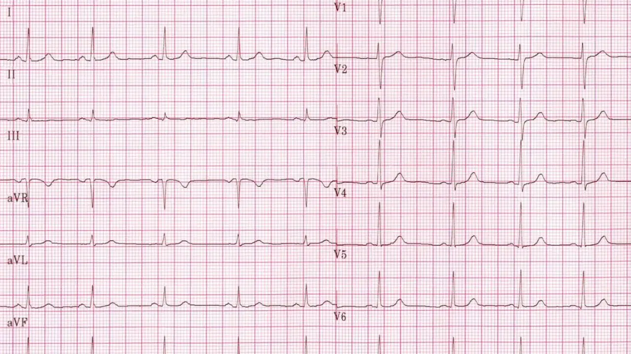
Interpretation of Results Normal results would include a heart rate between 60 and 100 beats per minute and a steady, regular heart rhythm.
What are the normal values of an electrocardiogram?
How to know if an electrocardiogram is good?
To interpret an electrocardiogram, it is necessary to assess the presence of these waves, their shape and duration, as well as the ST segment (time that elapses between the end of depolarization and the beginning of repolarization of the ventricles, measuring less than 1 mm , if greater than 1 mm indicates infarction or ischemia).
What does a bad EKG look like?
When this tracing has different shapes, it is considered that there is an abnormal electrocardiogram. However, this just means that there can be cardiac arrhythmias caused by bradycardia, a slow heart rate, or tachycardia, an increased heart rate.
How to know if an electrocardiogram is good?
To interpret an electrocardiogram, it is necessary to assess the presence of these waves, their shape and duration, as well as the ST segment (time that elapses between the end of depolarization and the beginning of repolarization of the ventricles, measuring less than 1 mm , if greater than 1 mm indicates infarction or ischemia).
What does a heart attack look like on an EKG?
The electrocardiographic diagnosis of AMI is based on the presence of ST-segment elevation >1 mm in two contiguous leads, or >2 mm in leads V1 to V4, or the appearance of complete left bundle branch block (LBBB). new.
What is the heart rate of a normal person?
Normally, the heart beats between 60 and 100 times per minute. In people who exercise regularly or take drugs to slow the heart rate, the heart rate may drop below 60 beats per minute.
What does V1 V2 V3 V4 V5 V6 mean?
THE THREE DIMENSIONS OF THE HEART IN THE ELECTROCARDIOGRAM In conclusion: V1 and V2 explore the septal area. V3 and V4 explore the anterior zone. V5 and V6 explore the lateral zone, together with I and aVL.
What does sinus rhythm mean?
Sinus rhythm is a term used in medicine to describe the normal heartbeat as measured on an electrocardiogram. It has some generic characteristics that serve as a contrast for comparison with normal electrocardiograms.
What does negative QRS mean?
When the QRS complex is clearly positive, it means that the electrical impulse approaches the measurement lead, if the QRS complex is negative, the impulse moves away from said lead, and an isobiphasic QRS complex means that the direction of the impulse is perpendicular to the lead. .
How to detect an arrhythmia on an electrocardiogram?
Electrocardiogram (ECG): is the simplest and most effective test to diagnose arrhythmias. It consists of recording the electrical currents of the heart through the placement of electrodes fixed to the patient’s skin, which allows analyzing possible alterations.
What is heart arrhythmia?
An arrhythmia is a disturbance of the heart rhythm. This is divided into two phases: diastole, the heart muscle relaxes and the cavity fills with blood, and systole, the muscle contracts and expels blood into the bloodstream, maintaining blood flow and blood pressure.
What does V1 V2 V3 V4 V5 V6 mean?
THE THREE DIMENSIONS OF THE HEART IN THE ELECTROCARDIOGRAM In conclusion: V1 and V2 explore the septal area. V3 and V4 explore the anterior zone. V5 and V6 explore the lateral zone, together with I and aVL.
What does the QRS wave on the electrocardiogram mean?
The QRS complex represents the depolarization that precedes contraction of the ventricles. A vector is used to represent the direction of electrical activity.
How to know if an electrocardiogram is good?
To interpret an electrocardiogram, it is necessary to assess the presence of these waves, their shape and duration, as well as the ST segment (time that elapses between the end of depolarization and the beginning of repolarization of the ventricles, measuring less than 1 mm , if greater than 1 mm indicates infarction or ischemia).
How do I know if I had a silent heart attack?
The only way to identify a silent infarction is imaging tests, such as an electrocardiogram or echocardiogram. If you think you’ve had a silent heart attack, talk to your doctor.
What if I have 80 beats per minute?
«On average, people who have 80 beats per minute at rest have a 30% higher risk of dying over the next 10 years compared to people who have 70 beats per minute,» says Dr. Albert Clará, head of the Department of Angiology and Vascular Surgery at Hospital del Mar and study signatory.
What should not be done before an electrocardiogram?
Preparing for the Test Do not exercise or drink cold water immediately before an ECG as these actions may cause false results.
What is V3 on an ECG?
V3: transitional lead between the left and right ECG potentials, as the electrode is in the interventricular septum. The R wave and S wave are usually about the same (isobiphasic QRS complex). V4: the lead of this lead is over the apex of the left ventricle, where the thickness is greatest.
When is the heart rate low?
The heart of adults at rest usually beats between 60 and 100 times per minute. If you have bradycardia, your heart beats less than 60 times a minute. Bradycardia can be a serious problem if your heart rate is too slow and your heart is unable to pump oxygen-rich blood around your body.
When to worry about an arrhythmia?
When to See a Doctor Seek immediate medical help if you experience shortness of breath, weakness, lightheadedness, lightheadedness, fainting or dizziness, and chest pain or discomfort. A type of arrhythmia called ventricular fibrillation can cause a dramatic drop in blood pressure.
What are benign arrhythmias?
Benign, which do not compromise the individual’s life, but have different medical management. Among them we can highlight, as they are the most common: – Extrasystoles, which are early heartbeats and usually do not require treatment, unless they cause a lot of discomfort.
How do you know if the QRS is positive or negative?
5.2 Calculation of the cardiac axis in degrees. That is, it is positive or negative when it is parallel to the lead we are looking at and that the positive QRS complex indicates that it is approaching and negative that it is moving away from this lead, but it will be isophase when the direction of the axis is perpendicular to the advance that we are observing .
What does QRS complex 80 mean?
The QRS complex is the graphic representation of the depolarization of the ventricles of the heart forming a pointed structure on the electrocardiogram. The QRS complex appears after the P wave, and because the ventricles have more mass than the cardiac atria, the QRS complex is larger than the P wave.
What is the abnormal T wave on an electrocardiogram?
The presence of negative T waves in the anterior precordial leads (V1-V4) in athletes is considered an abnormal finding and requires further studies to exclude the presence of an underlying cardiomyopathy, particularly HCM (where up to 2-4% of patients have waves. ..
What is the best test to see the heart?
A heart angiogram or angiogram is a procedure that uses contrast dyes and x-rays to look inside the arteries. It can show whether there is plaque blocking the arteries and the severity of the problem.